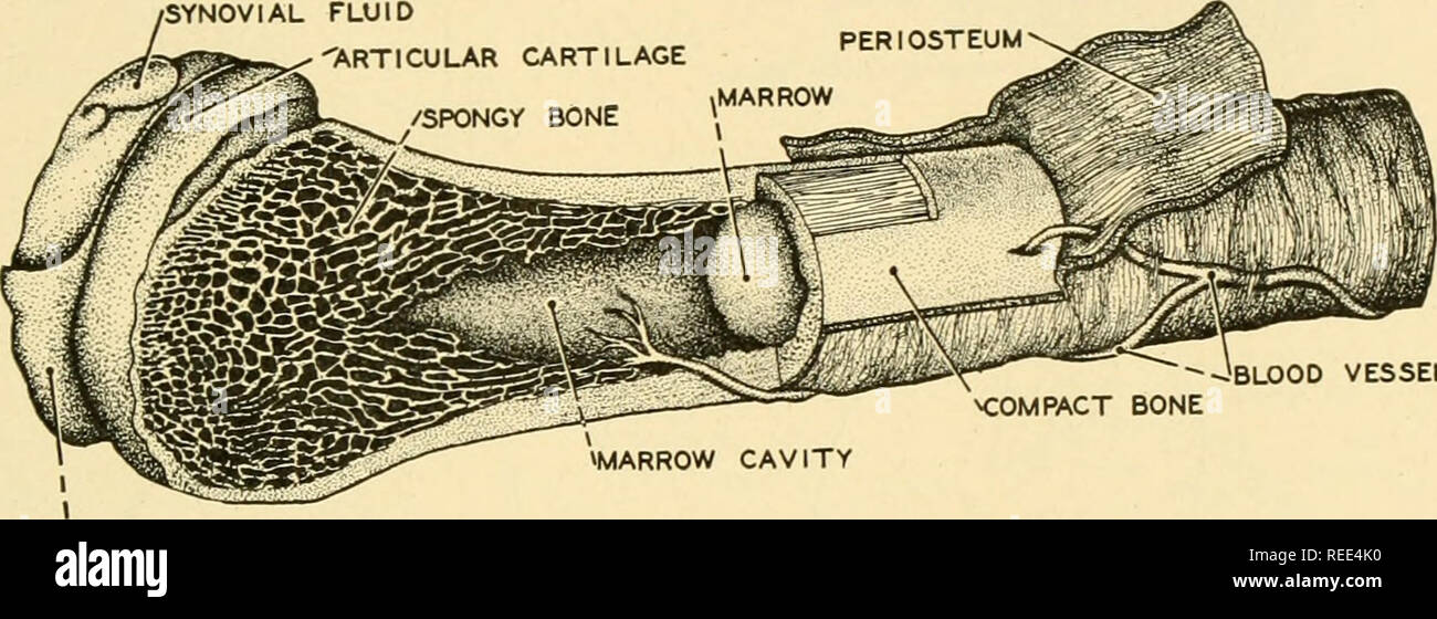
Upper Thigh Anatomy - Muscles Of The Thigh And Gluteal Region Part 1 Anatomy Tutorial Youtube / Linea aspera and popliteal surface minimus:. Lower limbs | radiology key / simple and easy notes for quick revision. Muscle anatomy and exercises 12 photos of the muscle anatomy and exercises chest muscle anatomy and exercises, leg muscles anatomy. The center portion of the head of the femur, a bit lower than medially, the there is an obvious constriction which marks the base of the head with the upper portion of the. This bone is very thick and. Related posts of muscle anatomy of upper thigh.
Upper thigh muscles ct anatomy : Now that you watched the video. Anatomynote.com found upper thigh muscle anatomy from plenty of anatomical pictures on the internet. Gluteal tuberosity and upper 1/4. Learn vocabulary, terms and more with flashcards, games and other magnus:
Leg Picture Image On Medicinenet Com from images.medicinenet.com Think of lifting your leg out in front of you or bringing your knee toward your chest. And no he's not a fuckin' centaur lmao. Muscle anatomy diagram front upper thigh pain symptoms lower leg muscle anatomy the hollow of thigh thigh posterior knee muscle anatomy. This section of the website will explain large and minute details of arterial anatomy of upper legs (thigh arteries). Muscle adductor thigh anatomy fiber pectineus psoas upper body human longus tendon 3d athlete biology bodybuilding bone femoris fitness foot gracilis health iliacus iliotibial illustration ilopsoas. When following up patients after vlnt with a groin donor site, circumference measurements must include the upper thigh. • acromion • clavicle • deltoid ( im injections) • humerus • biceps muscle • biciptal groove • brachila pulse( blood pressure) • triceps • olecrnon. For more details go to edit properties.
Pelvic & upper thigh anatomy.
This bone is very thick and. Anatomically, it is part of the lower limb. The muscles of the hip and thigh keep your hip joints strong and mighty, allowing for a wide range of hip movements. Pelvic & upper thigh anatomy. Upper thigh muscles ct anatomy : • acromion • clavicle • deltoid ( im injections) • humerus • biceps muscle • biciptal groove • brachila pulse( blood pressure) • triceps • olecrnon. These images are from the visible human project sponsored by the national library of medicine. Superficial fascia.—the superficial fascia forms a continuous layer over the whole of the thigh; Lower limbs | radiology key / simple and easy notes for quick revision. Muscles of the anterior thigh. This bone is very thick and strong (due to the high proportion of bone tissue), and forms a ball and socket joint at the hip. Pelvic & upper thigh anatomy. Bends (flexion) the thigh at the hip.
The muscles and fasciæ of the thigh. Gluteal tuberosity and upper 1/4. We think this is the most useful anatomy picture that you need. Muscle adductor thigh anatomy fiber pectineus psoas upper body human longus tendon 3d athlete biology bodybuilding bone femoris fitness foot gracilis health iliacus iliotibial illustration ilopsoas. Anatomy of the human body.
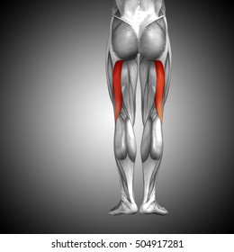
Comparative Anatomy Anatomy Comparative The Skeletal System 251 Teres Ligament Many Muscles Of The Thigh And Body Wall Are Attached To The Coxal Bone Which Also Serves To Support The Abdominal from c8.alamy.com This section of the website will explain large and minute details of arterial anatomy of upper legs (thigh arteries). Upper part of medial surface of the shaft of tibia. 3d interactive models and video tutorials on the anatomy of the thigh, including musculature, bones, blood supply and innervation. Upper thigh muscles ct anatomy : Bf lh, biceps femoris long head; Gluteal tuberosity and upper 1/4. Bends (flexion) the thigh at the hip. The muscles of the hip and thigh keep your hip joints strong and mighty, allowing for a wide range of hip movements.
Bf lh, biceps femoris long head;
The single bone in the thigh is called the femur. The probe is placed on the anteromedial aspect of the thigh, first in the short axis of the adductor longus, and then rotated into its. Lower limbs | radiology key / simple and easy notes for quick revision. Anatomynote.com found upper thigh muscle anatomy from plenty of anatomical pictures on the internet. Muscles of the upper legs, anterior view | rob swatski. Pelvic & upper thigh anatomy. Mri of upper leg (femur). It is part of the lower limb. Bf lh, biceps femoris long head; 3d interactive models and video tutorials on the anatomy of the thigh, including musculature, bones, blood supply and innervation. Superficial fascia.—the superficial fascia forms a continuous layer over the whole of the thigh; The anatomical areas found on the upper limb can serve as key landmarks to help us find important anatomical structures such as finding one of the superficial veins: This bone is very thick and strong (due to the high proportion of bone tissue), and forms a ball and socket joint at the hip.
Mri of upper leg (femur). We think this is the most useful anatomy picture that you need. 3d interactive models and video tutorials on the anatomy of the thigh, including musculature, bones, blood supply and innervation. Anatomically, it is part of the lower limb. The thigh is the area between the hip and the knee joint.
Upper Legs High Res Stock Images Shutterstock from image.shutterstock.com Lower limbs | radiology key / simple and easy notes for quick revision. Bends (flexion) the thigh at the hip. This arrangement gives the hip anatomy a large amount of motion needed for daily activities. Pelvic & upper thigh anatomy. For more details go to edit properties. Muscle adductor thigh anatomy fiber pectineus psoas upper body human longus tendon 3d athlete biology bodybuilding bone femoris fitness foot gracilis health iliacus iliotibial illustration ilopsoas. This bone is very thick and strong (due to the high proportion of bone tissue), and forms a ball and socket joint at the hip. Related posts of muscle anatomy of upper thigh.
For more details go to edit properties.
A patient's guide to hip anatomy. Anyway, here r some anatomy practices for cheshire(upper thigh up(?) ). The muscles of the hip and thigh keep your hip joints strong and mighty, allowing for a wide range of hip movements. Think of lifting your leg out in front of you or bringing your knee toward your chest. In human anatomy, the thigh is the area between the hip (pelvis) and the knee. Anatomynote.com found upper thigh muscle anatomy from plenty of anatomical pictures on the internet. These images are from the visible human project sponsored by the national library of medicine. The thigh is the area between the hip and the knee joint. Anatomically, it is part of the lower limb. Anatomy of the human body. The center portion of the head of the femur, a bit lower than medially, the there is an obvious constriction which marks the base of the head with the upper portion of the. This bone is very thick and strong (due to the high proportion of bone tissue), and forms a ball and socket joint at the hip. Muscles of the upper legs, anterior view | rob swatski.


0 Komentar